Before the Patient, the Cadaver: Inside a Hand and Wrist Arthroscopy Cadaveric Lab in Bangalore
Hand2Shoulder Clinic | Faculty note from the Arthrex cadaveric skills lab, Bangalore — 25 July 2026
There is a moment in every surgeon’s training that nobody photographs. It is the moment just before the first arthroscope goes into a live patient’s wrist — a joint the size of a walnut, packed with ligaments that are millimetres wide and unforgiving of error. Every surgeon remembers the feeling. Very few would say they felt ready.
The purpose of a cadaveric lab is to move that moment backwards in time. To make sure that when a young surgeon finally stands at the operating table with a real patient asleep in front of them, it is not the first time their hands have done that particular thing.
On 25 July, that is exactly what eight surgeons spent the day doing in Bangalore.
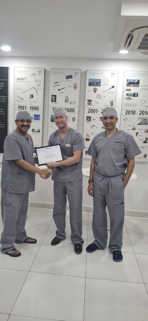
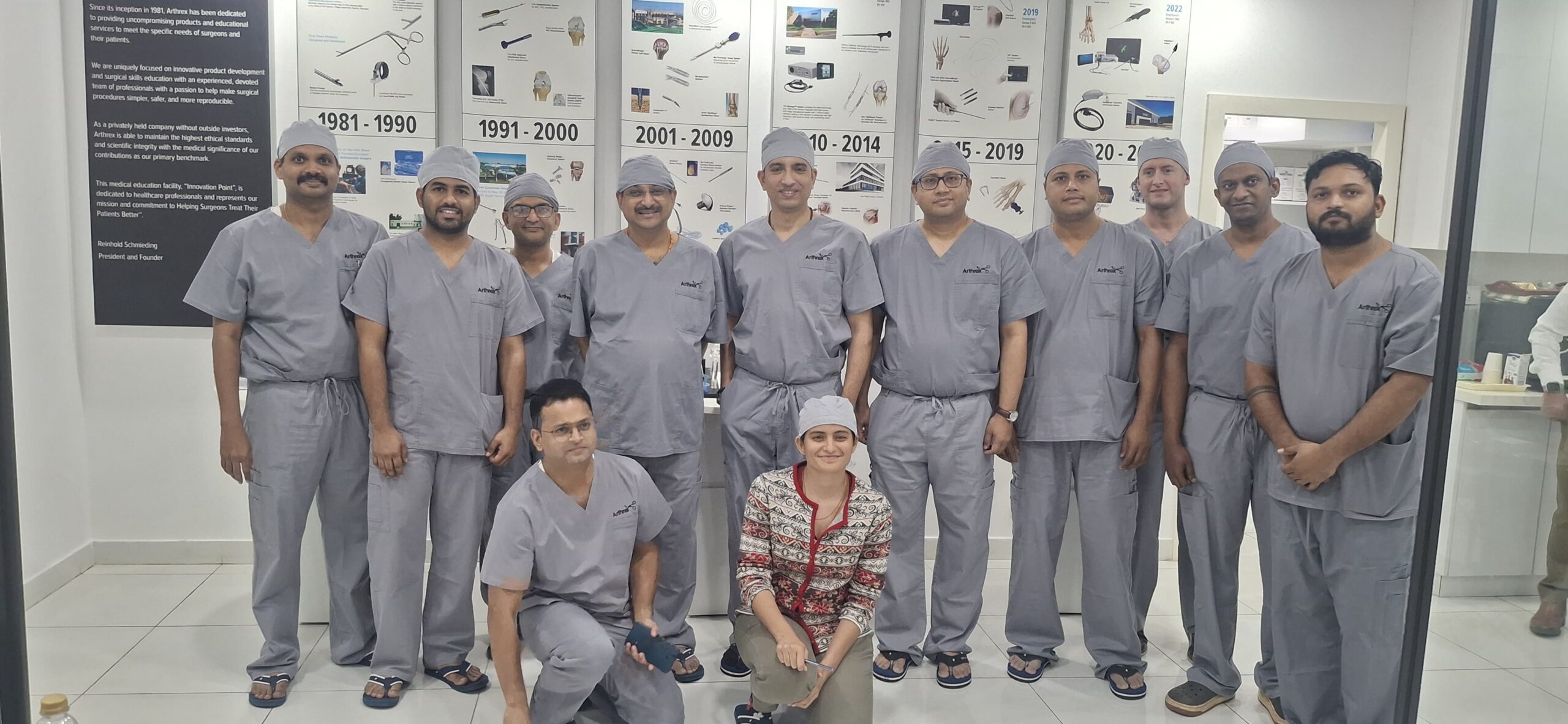
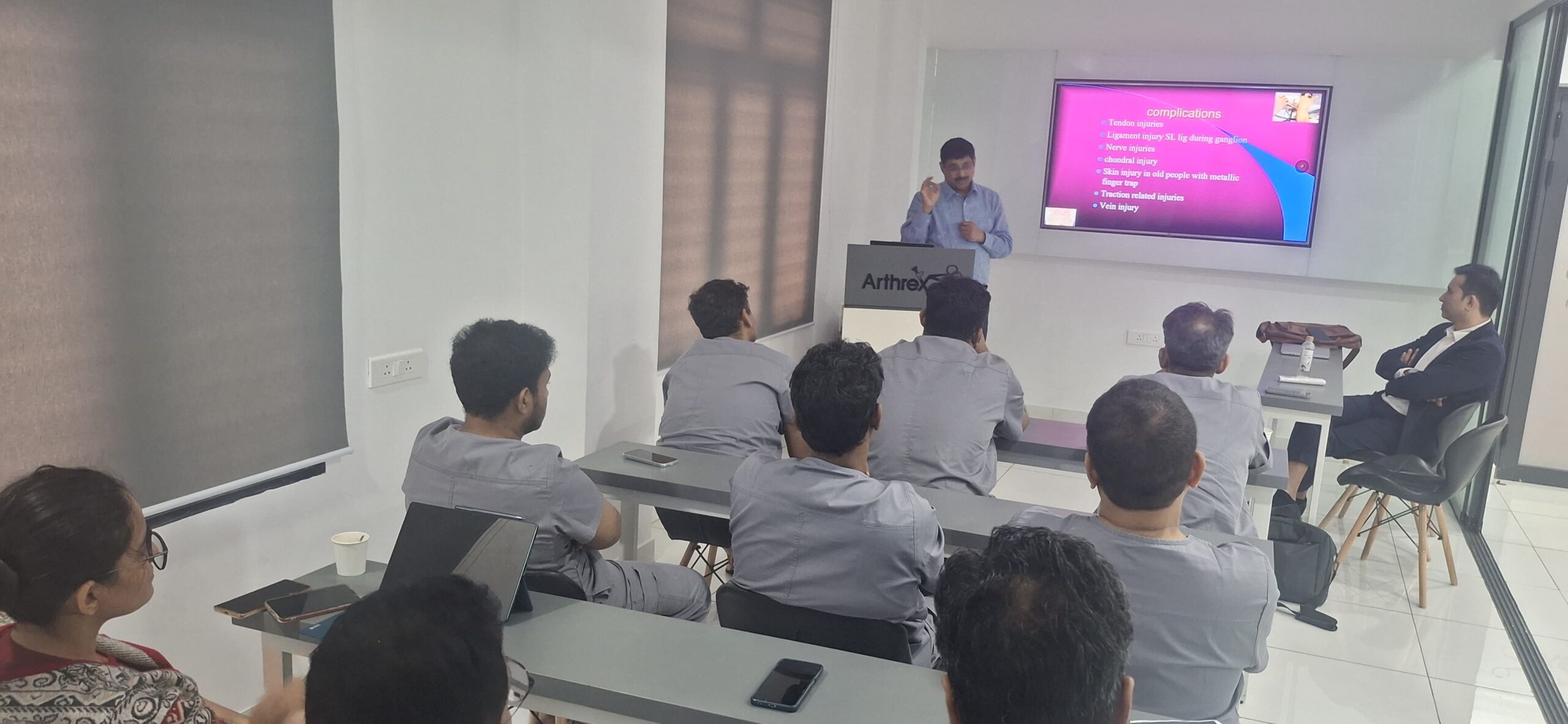
Eight Surgeons working at four cadaveric stations during a hand and wrist arthroscopy course in Bangalore. photo graphy in cadeveric was not permitted. Before going inside lab lectures explaining procedures were conducted.
An four-station lab, fully instrumented
This was not a demonstration where a room watches one pair of hands on a screen. Every participant had their own cadaveric specimen, their own arthroscopy tower, their own instrument set — camera, light source, shaver, radiofrequency probe, traction tower, suture anchors, suture passers and the full complement of small-joint arthroscopy instruments.
Four stations. Eight surgeons. Everyone scrubbed, everyone operating, nobody spectating.
That distinction matters more than it sounds. Arthroscopy of the wrist is not a visual skill, it is a proprioceptive one. Watching a triangulation manoeuvre on a monitor teaches you almost nothing about the wrist-and-elbow geometry your own arms need to find in order to bring an instrument to the tip of a 2.7 mm scope. You have to feel the angle fail, adjust, and feel it succeed. Repeatedly. That is only possible when the specimen in front of you is yours for the day.
The programme was built around four skill blocks, deliberately sequenced from foundation to reconstruction.
- Basic wrist arthroscopy and diagnostic survey
Everything begins here. Accurate portal placement — 3-4, 4-5, 6R, and the radial and ulnar midcarpal portals — is the single most under-taught step in wrist arthroscopy, and the single most common reason a case goes badly. We spent time on surface anatomy, on the safe interval relative to the extensor tendons and the dorsal sensory branch of the ulnar nerve, on establishing portals by nick-and-spread rather than blind stab, and on a disciplined, reproducible diagnostic sequence so that no compartment is ever missed.
Participants worked through the radiocarpal and midcarpal survey until the tour became automatic: scaphoid facet, radioscapholunate ligament, the scapholunate and lunotriquetral intervals, the articular disc, the ulnar recesses, and the midcarpal assessment of interosseous ligament competence. - Arthroscopic TFCC repair
The triangular fibrocartilage complex is the workhorse pathology of the ulnar-sided wrist, and it is where arthroscopy has changed practice most decisively. The station covered assessment of the disc — the trampoline and hook tests — classification of the tear pattern, and the technical differences between a peripheral capsular repair and a true foveal reattachment of the deep radioulnar fibres, which is what actually restores distal radioulnar joint stability.
Surgeons practised outside-in and all-inside repair techniques and worked with suture anchors for foveal fixation, including the knotless constructs that have made this repair considerably more reproducible than it was a decade ago. - Scapholunate ligament repair
Scapholunate injury is the most commonly missed significant wrist injury in India, and the price of missing it is high — a predictable march towards SLAC wrist arthritis over years. The station covered arthroscopic grading of instability, the dorsal component of the ligament as the structural priority, arthroscopic and arthroscopically-assisted repair with suture anchors, dorsal capsuloligamentous augmentation, and the decision points that separate a repairable acute injury from one that requires reconstruction.
We also discussed, frankly, the cases where repair is no longer the answer — because knowing when not to attempt a repair is as much a surgical skill as performing one. - Ulnar collateral ligament repair of the thumb
Skier’s thumb, gamekeeper’s thumb — whatever the eponym, an unrecognised complete UCL rupture at the metacarpophalangeal joint of the thumb costs a patient their pinch grip for life. The station covered clinical and radiological assessment of instability, identification of the Stener lesion (where the adductor aponeurosis blocks healing and makes non-operative treatment futile), anatomical footprint reattachment with suture anchors, and ligament augmentation techniques that permit early motion rather than six weeks in a spica.
Why cadaveric training is not optional
Hand and wrist surgery has a particular problem. The structures are small, the anatomy is dense, the margin for error is measured in millimetres, and the volume of any single procedure at any single centre is low. A surgeon may wait months between scapholunate cases. Under those conditions, the traditional model — see one, do one, teach one — simply does not deliver competence fast enough.
A cadaveric specimen offers something no simulator and no textbook can: real tissue planes, real ligament texture, real resistance under an instrument, and real consequences for a badly placed portal — without a single patient bearing the cost of the learning curve.
This is the entire motto of a lab like this one. The learning curve is real. The only question is who pays for it. In a cadaveric lab, the surgeon pays for it — in time, in concentration, in repetition. In the alternative model, the patient pays for it. There is no third option, and one of those two is not acceptable.
Passing it forward
Every technique in the modern hand surgeon’s repertoire — arthroscopic foveal repair, knotless anchor fixation, ligament augmentation, small-joint arthroscopy itself — was once new and once uncomfortable. It became routine only because someone took the trouble to teach it properly, hands-on, to the generation behind them.
That is the responsibility that comes with experience. Not to hold technique closely, but to hand it over in a setting where it can be learned safely and completely.
Eight surgeons left Bangalore on 25 July having done, with their own hands, procedures that many of them had until then only watched. Their future patients will never know that this day happened. That is precisely the point.